In the last week, we had a high number of corn leaf samples submitted to the U of I Plant Clinic. Most of are interested in knowing if their corn is infected with Goss's wilt, a bacterial disease showing up in many fields this growing season in Illinois. For more information on Goss's wilt, you can go to the following links:
Goss's wilt spreads across Illinois
Goss's wilt of Corn
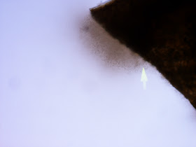
After a corn leaf sample is "checked-in" to the Plant Clinic by our awesome secretary, Deb, they are evaluated for disease. One of the first steps is to "check for ooze". If leaves are showing symptomology of a bacterial leaf disease, a cross-section is cut from the leaf tissue and examined under a microscope for bacterial streaming as seen in the picture below:
 |
| Bacterial ooze or streaming that can be seen under a microscope. |
If we see come bacterial streaming, we have immediately been testing the area showing symptoms, with an ELISA ImmunoStrip quick strip test for the bacteria,
Clavibacter, which is the Genus of the bacterial pathogen that causes the disease, Goss's wilt (
Clavibacter michiganensis)
. If the sample does not test positive for
Clavibacter, we assume that it is Stewart's wilt.
 |
| Corn leaf sample that has been found to show microscopic, bacterial streaming. |
 |
| Corn leaf tissue is ground up, put in the special bag with buffer solution, and a test strip is placed upright in the bag. |
 |
| Two red lines means that the corn sample was positive for Goss's wilt. | | | | | | | | |
Other corn leaf diseases that we have been finding are as follows:
 |
| Lesion of Common leaf rust (Puccinia sorghi) of corn. |
|
 |
| Common leaf rust (Puccinia sorghi) pustule as seen under a compound scope. Picture taken by Mike Meyer. |
|
 |
| Spores from Common leaf rust (Puccinia sorghi) as seen under a microscope. Picture taken by Mike Meyer. |
|
 |
| Gray leaf spot (Cercospora zeae-maydis) lesion. |
|
|
|
 |
| Gray leaf spot (Cercospora zeae-maydis) fruiting structures as seen under a compound scope. |
 |
| Gray leaf spot (Cercospora zeae-maydis) fruiting structure with spores as seen under a microscope. |
 |
| Cercospora zeae-maydis spores as seen under a microscope. |
 |
| Physoderma (Physoderma maydis) brown spot (Sorry no other spore pictures available at this time) |
|
|
Therefore, bacterial disease diagnosis is based on whether or not bacterial oozing is present as well as an ELISA ImmunoStrip quick strip test for
Clavibacter reads as positive or negative.
Fungal disease diagnosis are based on morphology of fungal fruiting structures and spores that are observed under a compound scope and microscope.


No comments:
Post a Comment
Note: Only a member of this blog may post a comment.