Friday, July 29, 2011

How We Identify Corn Leaf Diseases (Goss's wilt, Gray Leaf Spot, Common Rust and Physoderma brown spot) at the U of I Plant Clinic

In the last week, we had a high number of corn leaf samples submitted to the U of I Plant Clinic.  Most of are interested in knowing if their corn is infected with Goss's wilt, a bacterial disease showing up in many fields this growing season in Illinois.  For more information on Goss's wilt, you can go to the following links:
Goss's wilt spreads across Illinois
Goss's wilt of Corn

After a corn leaf sample is "checked-in" to the Plant Clinic by our awesome secretary, Deb, they are evaluated for disease.  One of the first steps is to "check for ooze".  If leaves are showing symptomology of a bacterial leaf disease, a cross-section is cut from the leaf tissue and examined under a microscope for bacterial streaming as seen in the picture below:
Bacterial ooze or streaming that can be seen under a microscope.
If we see come bacterial streaming, we have immediately been testing the area showing symptoms, with an ELISA ImmunoStrip quick strip test for the bacteria, Clavibacter, which is the Genus of the bacterial pathogen that causes the disease, Goss's wilt (Clavibacter michiganensis).  If the sample does not test positive for Clavibacter, we assume that it is Stewart's wilt.
Corn leaf sample that has been found to show microscopic, bacterial streaming.
Corn leaf tissue is ground up, put in the special bag with buffer solution, and a test strip is placed upright in the bag.
Two red lines means that the corn sample was positive for Goss's wilt.
Other corn leaf diseases that we have been finding are as follows:  
Lesion of Common leaf rust (Puccinia sorghi) of corn.
Common leaf rust (Puccinia sorghi) pustule as seen under a compound scope.  Picture taken by Mike Meyer.
Spores from Common leaf rust (Puccinia sorghi) as seen under a microscope.  Picture taken by Mike Meyer.
Gray leaf spot (Cercospora zeae-maydis) lesion.


Gray leaf spot (Cercospora zeae-maydis fruiting structures as seen under a compound scope.
Gray leaf spot (Cercospora zeae-maydis) fruiting structure with spores as seen under a microscope.
Cercospora zeae-maydis spores as seen under a microscope.
Physoderma (Physoderma maydis) brown spot (Sorry no other spore pictures available at this time)

Therefore, bacterial disease diagnosis is based on whether or not bacterial oozing is present as well as an ELISA ImmunoStrip quick strip test for Clavibacter reads as positive or negative.

Fungal disease diagnosis are based on morphology of fungal fruiting structures and spores that are observed under a compound scope and microscope.

Thursday, July 21, 2011

Where are the Cicadas Now? An Update on the Great Southern Brood

By: Jean Burridge, 
Certified Illinois Arborist, and U of I Plant Clinic Volunteer
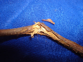
This has been an emergence year for periodical cicada in much of the southern part of the state. (See Phil Nixon's article in Home, Yard, and Garden Pest Newsletter, Issue 2). After thirteen years of subterranean existence, adults surfaced in late spring. Males sang, females listened; eggs were laid. Female cicadas possess a saw-like ovipositor, which they use to cut slits in twigs and small branches. They then insert their eggs into the wounds. The eggs will hatch in late summer, and the nymphs will drop to the ground and disappear into the earth. By August, an aggregation of insects so large and impressive that it has its own name (like Ann Arbor) will exist only inside the twigs of trees. Magicicada, indeed.

This feat of compression is not without cost. If you live in an area that is home to the brood, you may be seeing damage to trees, shrubs, or even perennials. Large trees look bedraggled – a bad hair year about sums it up. Trees that have been badly hit will have a fringe of dangling twigs. Weakened branches may snap immediately, or hang on until wind or ice loading finishes them off. 


The loss of apical dominance in an injured branch results in bushy unbalanced growth, as a host of lateral buds vie to replace the lost leader. The damage – broken branches, oviposition scars and aberrant growth – will be visible for years. 


It is too late to protect trees at this time. Native trees have co-evolved with cicadas – large trees usually grow out of their injuries and are not permanently harmed. Anything you can do to relieve stress would be helpful; water during dry spells and fertilize in the fall. 

Small trees or shrubs can succumb to this death of a thousand cuts; they may need to be replaced. If an emergence is anticipated, it is best to delay planting new trees until ovipositon has ceased.

Biologists suspect that periodical cicadas may have evolved to emerge on a thirteen or seventeen year cycle, in part because these are prime numbers. (Such protracted phenomena are very difficult for biochronologists to isolate – see Zivkovik. Mathematicians have modeled cicada broods – see Lehmann-Ziebarth et al.) Cicadas on this schedule can evade their predators, whose reproductive cycle cannot track them effectively. Animals who grew fat on cicadas have offspring who partake of no such abundance. When the brood reappears, there are no extra mouths to greet it. There is one animal, however – Homo sapiens - that is fully capable of exploiting a thirteen year interval. The Great Southern Brood will rise from the earth in 2024; be prepared….

Lehmann-Ziebarth, Nicholas , et al. “Evolution of Periodicity in Periodical Cicadas”. Ecology 86 (12) 2005. pp 3200-3211. http://www.math.wisc.edu/~milewski/Paul_Milewski,_Professor_of_Mathematics/Publications_files/cicadas.pdf  Web. 7/14/2011.
Nixon, Phil. “Periodical Cicada”. Home, Yard and Garden Pest Report Issue 2, May 2 2011. http://hyg.ipm.illinois.edu/article.php?id=263 Web. 7/14/2011
Zivkovik, Bora. “Centuries to Solve the Secrets of Cicadas”. http:www.scientificamerican.com/blog/post.cfm?id=too-hard-for-science-bora-zivkovil-2011-05-16. Web. 7/14/2011

Friday, July 15, 2011

The Corpse Flower is Blooming at the U of I Conservatory!

Excitement has been in the air at the U of I Plant Biology Conservatory as they anxiously await the bloom of the Titan arum or Corpse Flower.  It is 10 years old and is blooming for the very, first time right now!  It also is expected to be stinky, when the bloom opens!  The U of I Plant Biology Conservatory has recently extended it's hours for the public to see this flower. 

If you are unable to visit, you can see it on the live webcam !

Everyone at the U of I Plant Clinic was very excited about the bloom of this stinky flower.  In fact, we made sure the U of I Plant Clinic was "in on the action", by sending one of our students to visit the flower at the U of I Conservatory and wave to us from the webcam (Oh, yeah........ and put up a sign with a message from the U of I Plant Clinic)!
The "UI Plant Clinic "hearts" Titan"

The Titan and other plants can be seen at the U of I Plant Biology Greenhouse

Pictures of the Titan arum can be seen on the U of I Conservatory's Facebook Page

For more information, check out this fact sheet at U of I Plant Biology Greenhouse Plant Highlights


Press Releases:

Rare 'corpse flower' about to bloom at U of I Greenhouse

Rare Flower Expected to Bloom at UI

Seeds of Collaboration: Illinois Titan Arum Traces Roots to UW

Friday, July 8, 2011

A Few Tomato Diseases Seen at the U of I Plant Clinic this Week and How to Manage Them.


We confirmed 2 tomato samples from Cook County Illinois with TMV (Tobacco Mosaic Virus).  More information about this disease can be found at:  TMV - UMN.edu

How to manage TMV:
Rouge out the affected plants and sterilize pruning equipment. The virus is soil-borne, so crop rotation should be used to reduce the levels of inoculum. Avoid planting tomatoes, potatoes, peppers, and other solanaceous crops in the affected area for several years. Control solanaceous weeds, including jimpson weed as well as nightshade. Resistant varieties of tomato are available.


Septoria leaf blight is one of the most common diseases of tomato.  I have also two samples of tomatoes infected with this disease this week.  A University of Illinois publication about this and other tomato leaf diseases can be found at Tomato leaf diseases - UIUC.edu.  

How to manage Septoria leaf blight:
Due to the wet weather earlier this season, Septoria leaf spot has been particularly severe. To control Septoria leaf spot in the current growing season, remove infected plant parts and consider spraying weekly with an approved fungicide (or just accept losses). Be sure to avoid overhead irrigation which spreads disease, and stake plants to improve air circulation within the canopy. At the end of this season destroy all plant material. Rotate to a non-host crop next year. Products containing chlorothalonil (Bravo or Terranil, or others), copper, mancozeb (Dithane or pencozeb), or maneb are registered for use on tomato. Be sure to abide by the label you choose, especially the days between spraying and harvest.  Additionally, saving seed from season to season may increase disease because seed infection is possible with Septoria lycopersici.

Friday, July 1, 2011

Frogeye leaf spot or Phyllosticta leaf spot in Soybean? Easily confused?

A few weeks ago, there was an email floating around from "Ag Industry" that was claiming that frogeye leafspot, Cercospora sojina, had been found in soybeans.  This just did not seem possible to me, especially on leaves, from early vegetative growth stages on soybean, but never say never.

Several hundred plant samples later, I had forgotten about the frogeye leaf spot claim, until I was looking at a sample of 20 soybean leaves that had been collected from the Soybean Rust Sentinel Plot in Richland County, Illinois.


Then, the memory came flooding back to me when my Graduate Student asked me, "Is this frogeye leafspot?" as she leaned over my shoulder, while I was carefully looking at each leaf from the Richland Soybean Sentinel Plot under the dissecting scope.  I said, "No, this is PHYLLOSTICTA LEAF SPOT", Phyllosticta sojicola,  as I pointed out to her that the lesions had black, round fruiting structures within necrotic lesions, which can be easily seen with some magnification.  Also seen at this link: APS: Phyllosticta leaf spot with pycnidia

But, I told her that the early infection of Phyllosticta leaf spot sure could look like the lesions of frogeye leaf spot!  Both of these diseases are easily distinguished under a scope. Phyllosticta lesions will eventually grow, as the disease develops, and become "V-shaped" within soybean veins.  Phyllosticia leaf spot is not usually considered to be a serious disease.

I told Dr. Carl Bradly, University of Illinois Crop Sciences, Field Crop Plant Pathology, Extension Specialist, this story and he agreed with me that these two diseases could be confused.  In fact, he reminded me that he had written an article about this very subject in the The Bulletin in 2007: Similar Symptoms on Soybean: Phyllosticta Leaf Spot Vs. Frogeye Leaf Spot

A lot of attention has recently been given to frogeye leafspot, since this pathogen has been found to have reduced sensitivity to fungicides in Tennessee.  For more information: http://bulletin.ipm.illinois.edu/article.php?id=1429


For this reason, it is probably a good idea that frogeye leaf spot is correctly diagnosed.